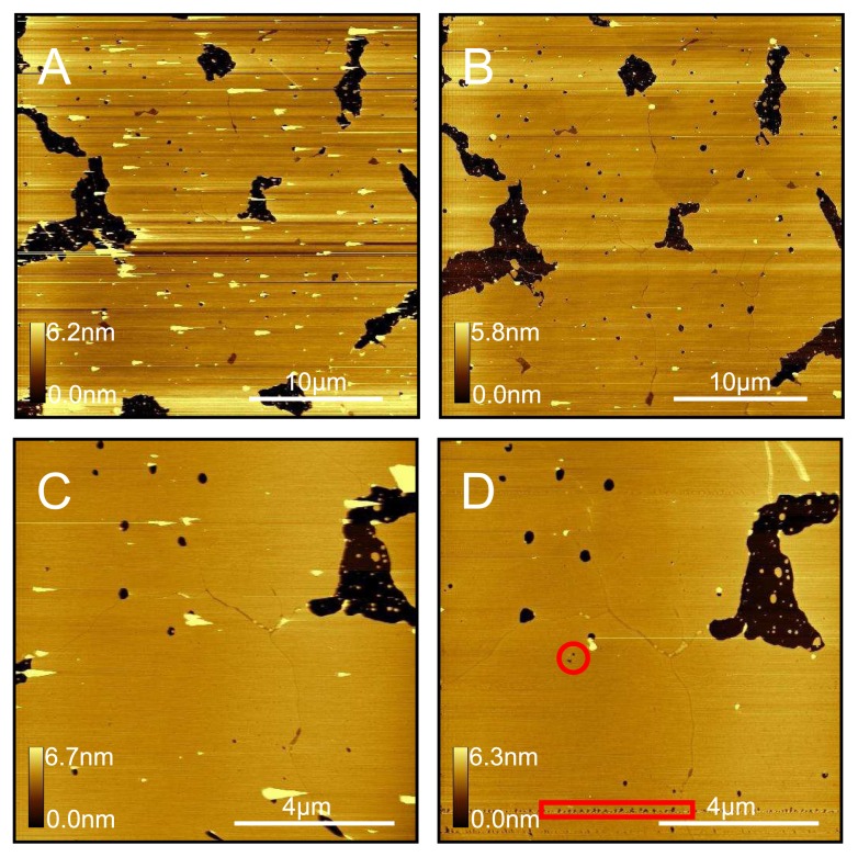
Figure 13.
DPPC sample prepared from HEPES-NaCl-Mg liposome solution and incubated at 60 °C for 60min. (A) Light force scanning highlights vesicles; (B) Higher force scanning makes the sample appear more continuous than it really is. Although some vesicles were dislodged and swept away, the smoother appearance of the surface is mostly due to the tip tracking across the vesicles better; (C) Extended regions of defect-free bilayer are observed; (D) A few lines of (C) were scanned with a high force and then scanned lightly again (area enclosed by red box). We see holes due to the hard scanning. We also see holes disappearing even with relatively light scanning (area enclosed by red circle), indicating that the DPPC bilayers are delicate and dynamic, although much less so than DOPC.

