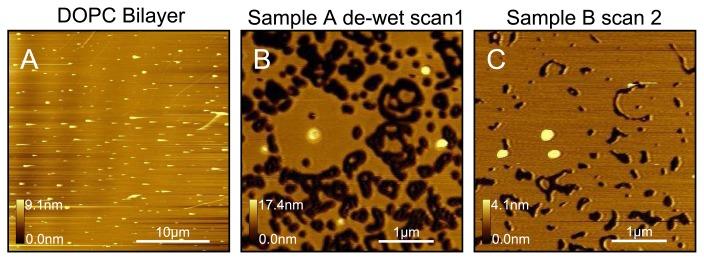
Figure 3.
Illustration of the dewetting test used to confirm bilayer coverage. (A) A sample is prepared using DOPC liposomes. Apart from some protrusions the sample is defect-free, making a confirmation of the state of the sample (mica, single bilayer, multilayer) difficult; (B) Sample instantaneously dewetted and then rehydrate. The holes confirm the presence of a single bilayer; (C) After a short period the holes begin to close, demonstrating again the fluidic nature of DOPC. All images were taken in pure water at room temperature.

