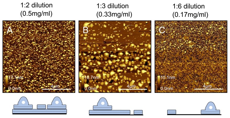
Figure 8.
Dilution test for samples prepared at 33 °C. DPPC samples in water were prepared at (A) 0.5mgml−1 ; (B) 0.33mgml−1 and (C) 0.17mgml−1 . By diluting the liposome solution to the point where bare mica is seen in the samples, we are able to see the very initial stages of vesicle fusion, which indicate protrusions are partially fused vesicles. Below: Schematics illustrating proposed generalised models for respective samples.

