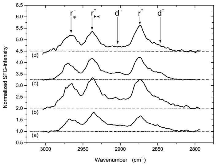
Figure 7.
HBM-FGF1 interaction as determined by SFS [171]. In situ SF spectra of (a) PG vesicle fusion; (b) rinsing of excess lipids; (c) equilibration with FGF1; (d) removal of FGF1, where r+ is the symmetric methyl stretch, rFR+ is the Fermi resonance of the symmetric methyl stretch, rip− is the in-plane methyl asymmetric stretch, d+ is the symmetric methylene stretch, and d− is the asymmetric methylene stretch.

