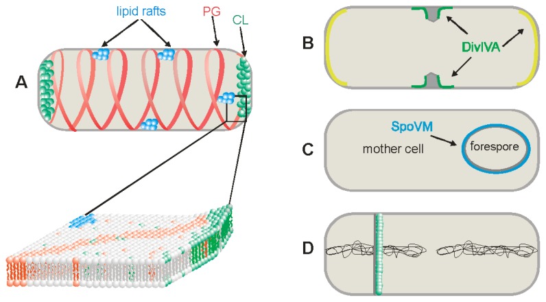
Figure 1.
Schematic presentation of different chemical and physical cues for protein localization into Bacillus subtilis membranes. (A) Presentation of three types of lipid domains—lipid rafts, cardiolipin (CL) and phosphatidylglycerol (PG) domains; (B) Negative curvatures membrane recognized by DivIVA. The highest negative curvature is near the forming septum, marked in green and moderate negative curvatures are at the poles, marked in yellow; (C) The positive curvature membrane, recognized by SpoVM, is located at the outer surface of the forespore in sporulating B. subtilis, marked in blue; (D) Asymmetrical de novo lipid synthesis in the sporulation septum from the mother cell side.

