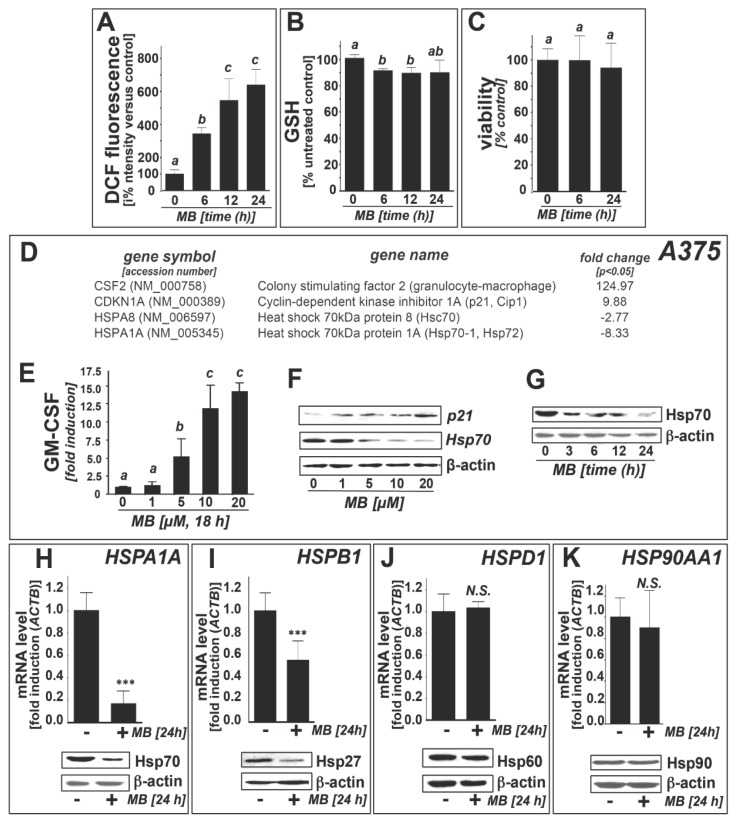
Figure 1.
Gene expression array analysis reveals heat shock response downregulation in human A375 melanoma cells exposed to methylene blue. (a) methylene blue (MB)-induction of cellular oxidative stress as monitored by DCF fluorescence (flow cytometric analysis; MB, 10 μM, ≤24 h exposure); (b) Depletion of reduced cellular glutathione; (c) Cell viability was examined using flow cytometric analysis (annexin V-PI staining). Numbers in the bar graph indicate viable (AV-negative, PI-negative) in percent of total gated cells (mean ± SD, n = 3); (d) gene expression changes by at least twofold (p < 0.05; RT2 Human Stress and Toxicity Profiler™ PCR Expression Array technology; MB: 10 μM, 24 h) in A375 melanoma cells. Arrays were performed in three independent repeats and analyzed using the two-sided Student’s t test; (e) MB-modulation (1–20 μM, 18 h; n = 3) of GM-CSF2 protein levels in A375 cell culture medium as assessed by ELISA; (f) MB-modulation (1–20 μM, 24 h) of Hsp70 and p21 expression (immunoblot analysis); (g) MB-modulation (10 μM, 3–24 h) of Hsp70 protein expression (immunoblot analysis); (h–k) Heat shock response gene expression changes (HSPA1A, HSPB1, HSPD1, HSP90AA1) at the mRNA and protein level induced by MB-treatment (10 μM, 24 h) in A375 cells using independent quantitative RT-PCR (upper panels) and immunoblot analysis (lower panels).

