
Fig 7.
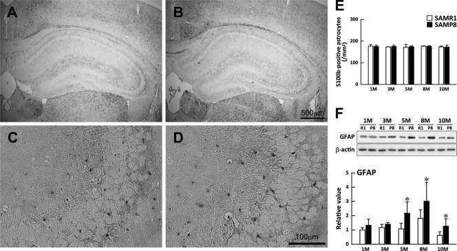
Astrocyte density and GFAP content of the SAMP8 and SAMR1 hippocampus at different ages. Astrocyte cell bodies were visualized by immunostaining with anti-S100B antibody in the whole hippocampus (A,B) and CA3 region (C,D) of SAMR1 (A,C) and SAMP8 (B,D) mice at 3 months. E: Number densities of astrocytes in the whole hippocampus of SAMR1 (open bars) and SAMP8 (solid bars) were obtained by counting S100B-positive cell bodies at different ages and expressed as mean + SD [n = 3–8 (SAMR1, SAMP8)]. F: GFAP content of the SAMR1 (open bars) and SAMP8 (solid bars) hippocampus quantified by Western blotting with anti-GFAP antibody. Immunoreactive bands of GFAP (examples shown in the upper panel) at different ages were quantified using β-actin as standard and expressed relative to that of SAMR1 at 1 month. Results are expressed as means + SD [n = 5–8 (SAMR1), n = 5–6 (SAMP8)]. *P < 0.05 between age-matched SAMP8 and SAMR1.