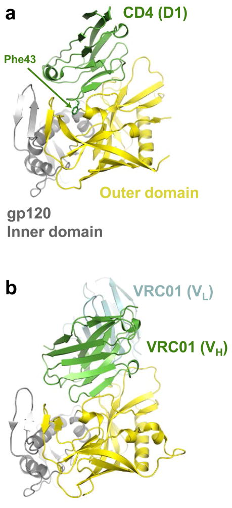
Figure 2.
CD4 and CD4-mimicking antibody binding to the gp120 core. (a) Structure of HIV-1 gp120 (outer domain is shown in yellow and inner domain in gray) in complex with CD4 (green; pdb code 3JWD). Only immunoglobulin-like domain 1 (D1) of CD4 is shown; the Phe43 side-chain is depicted as sticks. (b) VRC01 antibody–gp120 co-crystal structure (pdb code 3NGB, heavy chain shown in green and light chain in cyan) oriented as in panel a. Only the variable domains of the heavy (VH) and light (VL) chains of the antibody are shown.

