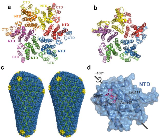
Figure 3.
HIV-1 capsid structures. Crystal structures of the hexameric (a, pdb code 3H47) and pentameric (b, pdb code 3P05) full-length HIV-1 CA assemblies. Individual subunits are coloured by chain. (c) The model for the complete HIV-1 capsid, based on the crystal structures 32. NTDs of the hexameric and pentameric CA units are shown in blue and yellow, respectively; CTDs are green. (d) HIV-1 CA NTD in complex with PF-3450074 (PF, pdb code 2XDE). The orientation is related to that of the blue NTD in panel a by an ~100° rotation, as shown. Residues critical forPF -3450074 binding as revealed by resistance mutations 44 are indicated.

