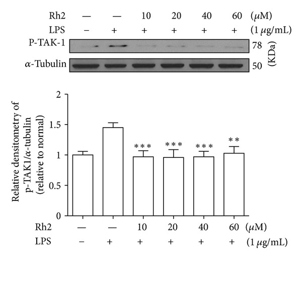
Figure 3.

Effects of ginsenoside Rh2 on TAK1 phosphorylation. RAW 264.7 cells were pretreated with indicated concentrations of ginsenoside Rh2 for 1 h prior to incubation of LPS (1 μg/mL) for 30 min. p-TAK1 was determined by western blot. Each immunoreactive band was digitized and expressed as a ratio of α-tubulin levels. The ratio of the normal group band was set to 1.00. Data are expressed as mean ± SD of three independent experiments. ***P < 0.001, significantly different when compared with LPS-stimulated cells.
