
Figure 2.
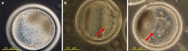
The location of PVP solution in embryos. a PVP was generally not observed in the embryo and appeared similar to the control. b PVP generally dispersed with some PVP remaining at the injection site. c Entire volume of PVP remaining in parts of the oocyte. Arrows show the localization of PVP solution. Figure from Kato and Nagao [30]