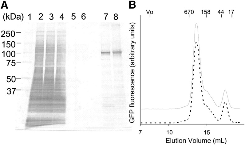
Fig. 3.
Purification of human ABCG1 expressed in FreeStyle 293-F cells. A: Silver staining of SDS/PAGE (5–20% gradient gel). Lane 1, size marker; lane 2, crude membranes; lane 3, crude membrane proteins solubilized with 0.8% DDM; lane 4, unbound proteins to FLAG agarose; lane 5, washout proteins with 0.8% DDM; lane 6, washout proteins with 0.05% DDM; lane 7, purified ABCG1-GFP (0.3 μg); lane 8, purified ABCG1(KM)-GFP (0.3 μg). Equal volumes of sample were loaded in lanes 2–6. B: FSEC analysis of the purified ABCG1-GFP (solid line) and ABCG1(KM)-GFP (dotted line).

