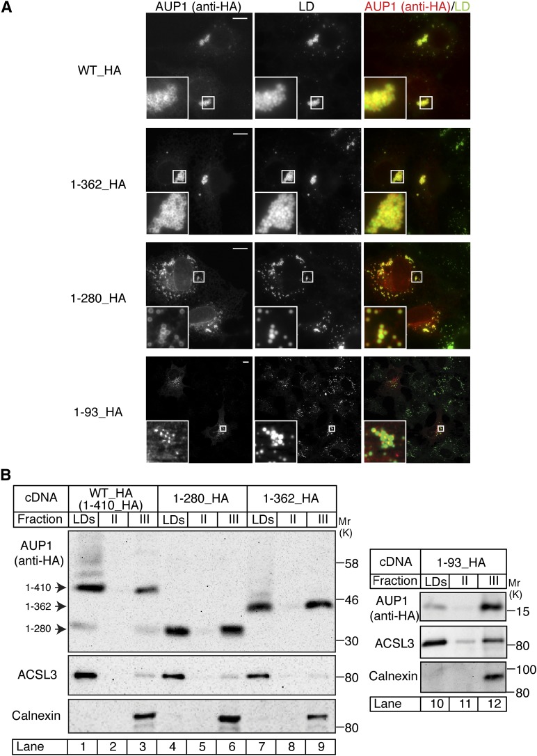
Fig. 2.
Identification of the lipid droplet targeting domain of AUP1. A: COS-7 cells expressing fusion proteins as indicated in Fig. 1A were fixed and processed for immunofluorescence microscopy using anti-HA antibody to detect the transfected AUP1 and the LD-specific dye LD540 to detect LDs. Scale bars, 10 μm. B: COS-7 cells expressing fusion proteins as indicated were grown in medium supplemented with 150 μM oleic acid to promote LD formation. LDs were isolated by floatation in a sucrose gradient. Proteins of the floating LD fraction (LDs) and the two lower fractions (II, III) were analyzed by SDS-PAGE/immunoblotting using antibodies against the transfected AUP1 (anti-HA), LD marker (ACSL3), and ER marker (calnexin).

