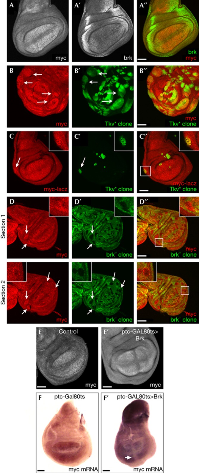
Figure 3.
Brk represses Myc expression in lateral regions of the wing disc. (A–A′′) Myc and Brk are expressed in complementary regions of the wing disc. Wild-type wing disc stained with anti-Myc (A,A′′) and anti-Brk (A′,A′′). (B–B′′) Clones expressing activated Thickveins (TkvQ235D) display elevated levels of Myc protein. Clones marked with green fluorescent protein (GFP) in green and with α-Myc in red. (C–C′′) Clones expressing activated Thickveins (TkvQ235D), marked by GFP (green), display elevated levels of Myc transcription, detected by a lacZ enhancer trap in the Myc locus (P{lacW}dmG0359 in red). Displayed clone is boxed in C′′. (D–D′′) Brk mutant clones (marked by the loss of GFP, green) have elevated levels of Myc protein (α-Myc, red) laterally (arrows) but not medially in the disc where Brk is not expressed. Two sections of the same disc are shown. Displayed clone is boxed in D′′. (E–E′′) Medial expression of Brk represses Myc protein accumulation. Upstream activating sequence (UAS)–Brk was transiently induced in a medial stripe using patched-GAL4, GAL80ts by shifting larvae grown at 19–29°C for 12 h causes reduced Myc protein levels (E′), not observed in control discs (E). (F–F′) Transient, medial expression of Brk, as in E, represses Myc mRNA accumulation, detected by in situ hybridization. Scale bar, 50 μm. Disc genotypes: (A–A′′) w1118 (B–B′′) hsFlp, Act>STOP>GAL4, UAS–GFP, UAS–TkvQ235D (C–C′′) Myc–lacZ(P{lacW}dmG0359), hsFlp, Act>STOP>GAL4, UAS–GFP, UAS–TkvQ235D (D–D′′) hsFlp, FRT brkXA/FRT GFP (E,F) ptc–GAL4, GAL80ts (E′,F′) ptc–GAL4, GAL80ts, UAS–brk. Brk, Brinker; ptc, patched.

