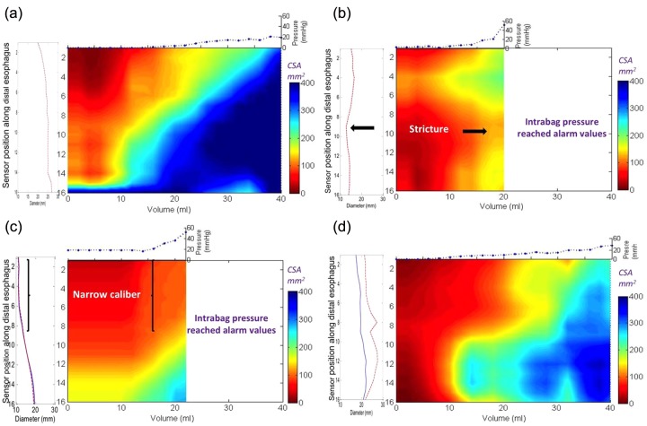
Figure 6.
Functional luminal imaging probe (FLIP) topography plots of the same three patients with eosinophilic esophagitis (EoE) and control as illustrated in Figure 5. Note that the detail of the regional changes can distinguish between the patients with EoE. The bag distends first in the distal esophagus due to dependent filling and increased distensibility in the distal esophagus in both the control (a) and the patients with EoE and a normal endoscopy (d). However, the intrabag pressures are higher in the patient with EoE at maximal volumetric distention. The regional changes in the patient with EoE and a narrow caliber (c) are illustrated by the long segment in the proximal measurement area where the cross-sectional area (CSA) does not rise above 150 mm2 despite much higher pressure values compared with the control subject. Similarly, the patient with a focal stricture (b) also maintains a persistent zone of reduced CSA in the middle measurement area despite the higher pressure values recorded in the bag. Both patients with EoE and a focal stricture and a narrow caliber reached intrabag pressures greater than 40 mmHg with smaller distention volumes compared with the control subject and the patient with EoE and normal endoscopy.

