
Figure 3. Comparison of thalamic segmentations.
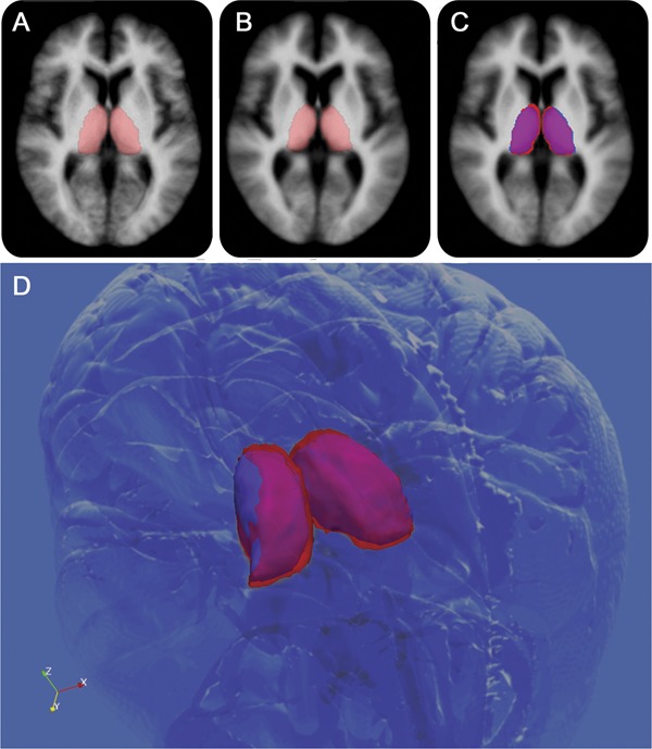
Comparison of thalamic segmentations between 26 healthy controls (HC) (age- and sex-matched) (A) and 98 patients with relapsing-remitting multiple sclerosis (MS) (B). (C) Voxel-wise differences between the groups, with magenta representing common areas, red HC-only areas, and blue MS-only areas. Individual patient images were coregistered into the Montreal Neurological Institute 152 space for visualization. A 3D view (D) shows that the MS thalami are smaller overall and slightly shifted away from the midline (most likely by global atrophic processes).