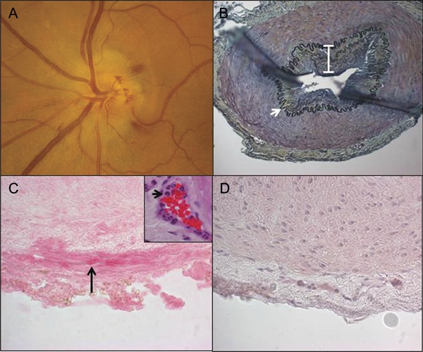
Figure. Fundus examination, histology, and immunohistochemistry on temporal artery biopsy of varicella-zoster virus ischemic optic neuropathy.

(A) Fundus photograph of the left eye reveals a swollen elevated optic disc with blurred margins and peripapillary flame hemorrhages. (B) Histologic and immunohistochemical analysis of the left temporal artery biopsy obtained 4 days after the patient's onset of loss of vision. Modified Movat pentachrome stain of a formalin-fixed paraffin-embedded section of the temporal artery demonstrates a thickened intima (white bar) and nearly intact internal elastic lamina (white arrow). Original magnification ×200. Note the presence of varicella-zoster virus (VZV) antigen in the adventitia of the temporal artery stained with anti-VZV antibody (C, black arrow, pink, ×600 magnification), but not with normal rabbit serum (D), and an abundance of neutrophils in the adventitia around the vaso vasorum vessels (C, inset, black arrow, hematoxylin & eosin, ×600 magnification).
