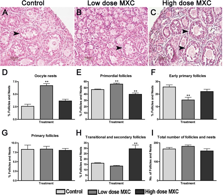
FIG. 1.
Effect of fetal and neonatal MXC exposure on in vivo follicle stage composition in postnatal day (PND) 7 ovaries. Representative images of hematoxylin and eosin-stained histology of 5 μm ovary sections from PND 7 rats exposed during fetal and neonatal periods to vehicle (DMSO:oil [1:2], Control; A), 20 μg/kg/day MXC (low-dose MXC; B), and 100 mg/kg/day MXC (high-dose MXC; C). Primordial through secondary follicles (arrowhead) were observed in control, low-dose MXC, and high-dose MXC, but with a predominant number of primordial follicles and oocyte nests (B) in the low-dose MXC and increased numbers of transitional and secondary follicles in the high-dose MXC group. Bar = 50 μm. Follicles and nests were counted from the two largest serial sections selected from the center of the ovary and classified into oocyte nests (D), primordial follicles (E), early primary follicles (F), primary follicles (G), and transitional/secondary follicles (H). The area of each cross-section was measured with an optical micrometer, and the total follicles counted were normalized to the area of each cross-section (I). The percentage of oocyte nests or each follicle type was calculated based on the total number of follicles and nests per entire ovarian section. In addition, the entire nest, rather than each oocyte within the nest, was counted as one; n = 5–7 ovaries (animals) from at least three litters per treatment group. Error bars represent SEM. **P < 0.01.

