
FIG. 2.
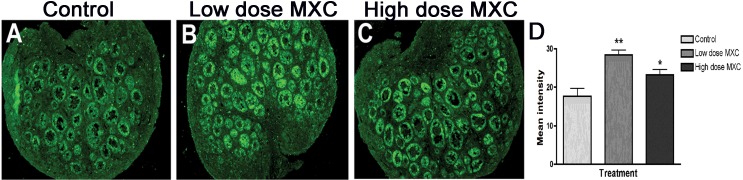
The effect of fetal and neonatal MXC exposure on AMH expression in PND 7 ovaries. Representative images from immunohistochemical analysis of AMH in PND 7 rat ovaries treated with vehicle, (DMSO:oil [1:2], Control; A), 20 μg/kg/day MXC (low-dose MXC; B), and 100 mg/kg/day MXC (high-dose MXC; C). Original magnification ×100. Quantification of AMH expression was performed on the whole ovary area using Image J software (NIH Image). There was a significant increase in the levels of AMH in low-dose MXC-treated and high-dose MXC-treated ovaries (D). Analysis was conducted with four separate animals per treatment in each experiment from at least three different litters. Error bars represent SEM. *P < 0.05 and **P < 0.01.