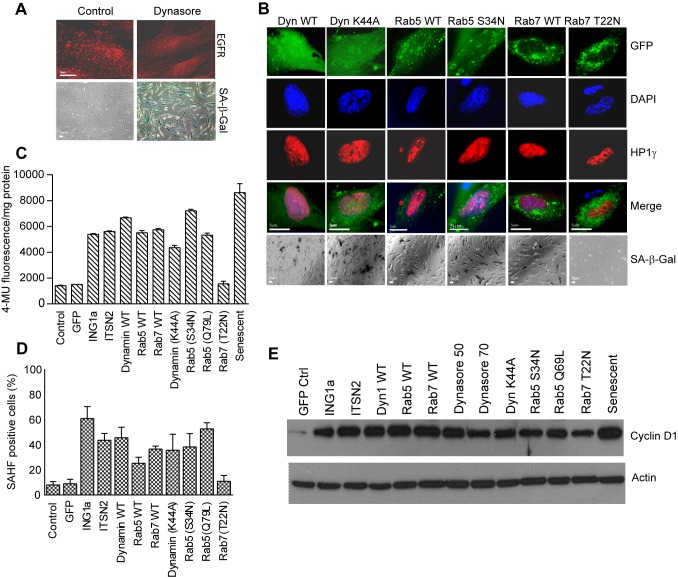
Figure 7. Inhibiting endocytosis leads to senescence.
(A) Cells treated for 48 h with 50 µM dynasore were fixed and stained for EGFR endocytosis. Lower panels show fields of cells similarly treated and stained for the presence of SA-β-gal. (B) Constructs encoding Dynamin1 (WT & K44A mutant), Rab5 (WT & S34N), or Rab7 (WT & T22N) were transfected into young primary fibroblasts, and 24 h later, cells were fixed and stained with DAPI to visualize DNA with anti-HP1γ to visualize SAHF and with X-gal (at pH 6.0) to identify cells with SA-β-gal activity. (C) SA-β-gal activity in transfected cells was quantified using methylumbulliferryl-β-D-galactopyranoside (MUG); values were normalized to the total protein concentration of each cell lysate estimated by Lowry assay, 24 h after transfection. (D) Quantification of cells positive for the presence of SAHF from Figure 7B is plotted as histogram by cell counting. (E) Cell lysates from low passage (28 MPD) Hs68 cells transfected with the indicated constructs were immunoblotted for the cyclin D1 senescence marker. Actin was used as the loading control.

