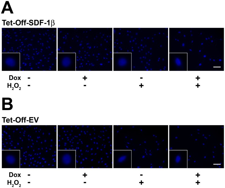
Figure 3. SDF-1β preserves BMSC nuclear morphology following exposure to H2O2.
Representative fluorescence micrographs of Hoechst 33342-stained A) Tet-Off-SDF-1β BMSCs and B) Tet-Off-EV control BMSCs after vehicle control or H2O2 treatment. Overexpression of SDF-1β in Tet-Off-SDF-1β BMSCs allows for a greater number of surviving cells and cells with preserved nuclear morphology after H2O2 treatment compared to Dox-suppressed and Tet-Off-EV controls (6 h, ±100 ng/ml Dox, ±1.0 mM H2O2, 20×, 40×, bar 100 µm, n = 3, 3 independent experiments).

