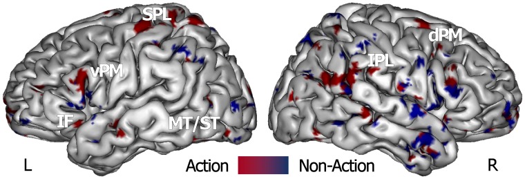
Figure 2. Map of the combined ‘supramodal’ SVM classifier that was defined by using the training data from all action (red scale) and non-action (blue scale) stimuli classes, and was employed in a ‘knock-out’ procedure.
Spatially normalized volumes are projected onto a single-subject inflated pial surface template in the Talairach-Tournoux standard space. Ventral and dorsal areas of the premotor cortex (vPM e dPM), inferior frontal (IF) cortex, superior and middle temporal gyri (ST/MT), superior (SPL) and inferior parietal lobule (IPL).

