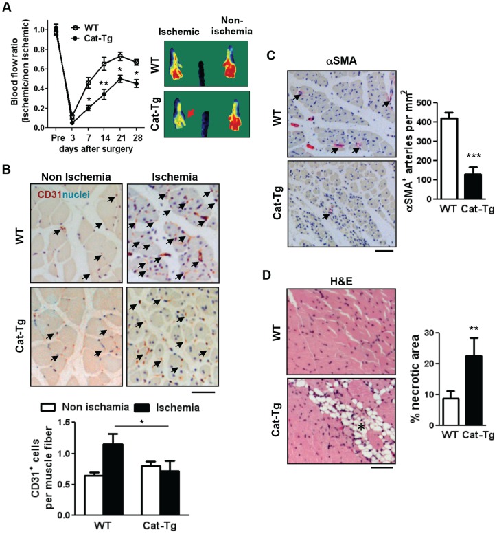
Figure 1. Endothelial catalase overexpression impairs post-ischemic neovascularization.
A, Wild-type (WT) and Tie2-driven catalase transgenic (Cat-Tg) mice were subjected to hindlimb ischemia. Blood flow recovery was measured by relative values of foot perfusion between ischemic and non-ischemic legs (WT n = 9, Cat-Tg n = 7,). Representative laser Doppler images at day 28 (right panels). B, capillary formation in the ischemic gastrocnemius muscles were analyzed by immunostaining with an endothelial-marker CD31 (red-brown and arrows) and quantified as capillaries per muscle fibers in the graph (n = 4 mice per group and bar = 50 μm). C, the density of arterioles are measured by αSMA staining (red and arrows) on the ischemic gastrocnemius muscles (n = 4 mice per group and bar = 50 μm). D, tissue repair after ischemic injury was examined in the ischemic gastrocnemius muscles with haematoxylin and eosin (H&E) staining which show necrotic regions with fibro-adipose tissue infiltration (asterisk in the image) as a sign of impaired or delayed repair process (n = 4 mice per group and bar = 50 μm). All data shown are mean+SE (*p<0.05 and **p<0.01).

