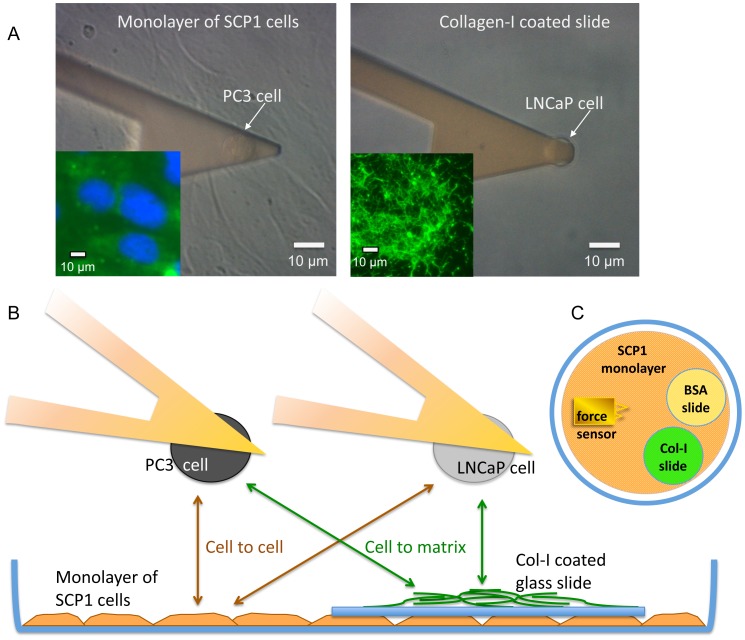
Figure 2. Schematic representation of the experimental setup.
(A) Phase contrast images of a prostate cancer cell attached to the cantilever (arrows) above an SCP1 monolayer (left) and a Col-I-coated slide (right). The scale bars indicate 10 µm. On the lower left corners immunofluorescence images are inserted. Col-I, labeled with AlexaFluor488 fluorescence dye appears in green and cell nuclei, stained with DAPI in blue. (B) Single cells from two different prostate cancer cell lines (PC3 and LNCaP) were immobilized to a tipless AFM cantilever (force sensor) in order to study their interaction forces with the apical surface of a SCP-1 monolayer (representing mesenchymal stem cells) or with Col-I (representing bone matrix). (C) Schematic top view of the culture dish lid with a BSA-coated glass cover slip (as substrate for fishing a gently injected prostate cancer cell) and a Col-I coated glass cover slip both on top of a monolayer of mesenchymal stem cells. For calibration and fishing a cell, the force sensor visits the BSA slide, for the experiment on collagen the Col-I slide and for the experiment on mesenchymal cells the SCP1 monolayer.

