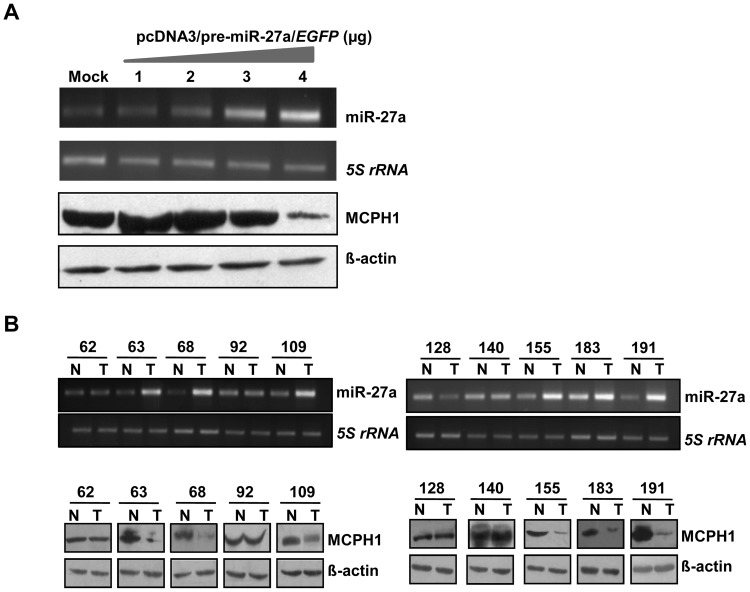
Figure 9. miR-27a negatively regulates MCPH1.
(A) miR-27a negatively regulates MCPH1 level in KB cells. Representative images of the correlative expression of MCPH1 protein level after the transient transfection of the pcDNA3/pre-miR-27a/EGFP construct in KB cells. Note the reduced expression of MCPH1 upon overexpression (4 µg) of pcDNA3/pre-miR-27a/EGFP. Mock lane represents transfection with the empty vector pcDNA3/EGFP. 5S rRNA and ß–actin were loading controls for RT-PCR and Western blotting respectively. (B) The correlative expression analysis of miR-27a and MCPH1 in 10 paired OSCC samples. Note a negative correlation between the levels of miR-27a and MCPH1 in six matched samples: 63, 68, 109, 155, 183 and 191. However, no correlation between the levels of miR-27a and MCPH1 was observed in three matched samples: 62, 92 and 140. In the remaining one matched sample (pt# 128), the level of miR-27a was downregulated in tumor, but the level of MCPH1 was unchanged in the tumor tissue. 5S rRNA and ß–actin were loading controls for RT-PCR and Western blotting respectively. Abbreviations: N, normal oral tissue; and, T, tumor oral tissue. The numbers refer to patient numbers.

