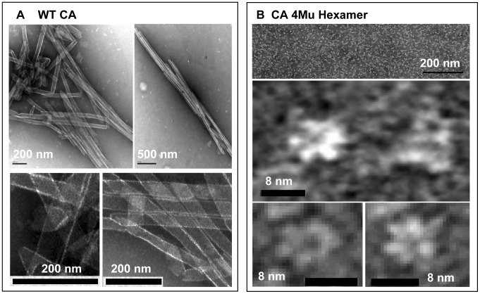
Figure 4. Transmission electron microscopy analysis of structures present in the solution of (A) polymerized WT CA and (B) assembled CA 4Mu Hexamer.
Objects were deposited onto the grid and visualized by the negative staining. Black bar on micrographs represents scale in nm. (A) Tube-like and cone-like structures can be observed in solution of WT CA polymerized at 40 µM concentration in buffer containing 50 mM sodium phosphate (pH 7.5), 2 M NaCl, 0.005% Antifoam 204. (B) Hexameric assemblies are clearly visible in a solution containing 19.5 µM of disulfide cross-linked CA 4Mu (expressed as CA monomers concentration) in 20 mM Tris (pH 8.0), 10% glycerol buffer.

