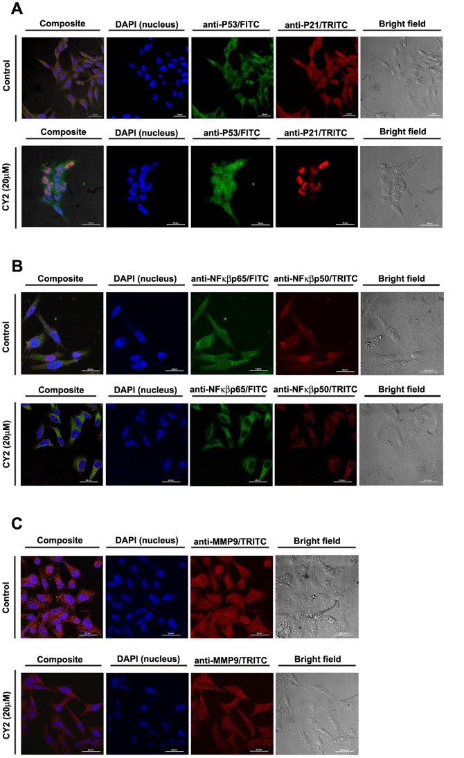
Figure 9. Immunocytochemistry showing localization of P53, P21, NF-κB p50, NF-κB p65 and MMP-9 in presence and absence of CY2 in HCT116 cells.
DAPI was used for genomic DNA counterstaining (A) Translocation status of P53 and P21 in control cells (upper panel) and 20 µM CY2 treated cells (lower panel). HCT116 cells coincubated with primary antibodies for P53 and P21 and developed with FITC (λEm∼525 nm, green) and TRITC (λEm∼576 nm, red) labeled secondary antibodies respectively, cotranslocation (left composite panel) in the nuclei was observed for both of these proteins with respect to the untreated cells, where they were found to reside mainly in the cytoplasm. Scale Bar = 20 µm. (B) Translocation status of NF-κB p50 and NF-κB p65 in control cells (upper panel) and 20 µ M CY2 treated cells (lower panel). The nuclear localization of the two main subunits of the NF-κB family viz. p65 and p50 (developed with FITC and TRITC -labeled secondary antibodies respectively) was found in untreated control cells but upon treatment, these two subunits tends to localize into the cytoplasm with respect to the untreated control as shown in the composite confocal micrograph. Scale Bar = 15 µm. (C) Localization of MMP-9 in control cells (upper panel) and 20 µM CY2 treated cells (lower panel). Confocal microscopy revealed that immunocytochemistry of MMP-9 protein (developed with TRITC-labelled secondary antibody) expression decreased following treatment with CY2 with respect to the untreated control. Scale Bar = 15 µm.

