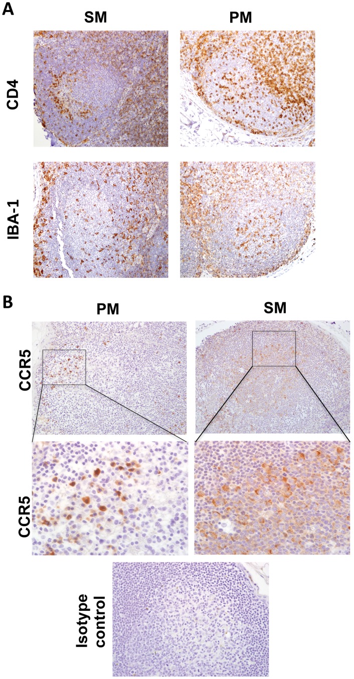
Figure 4. Immunophenotype of resident GC cells in SM and PM by IHC. A, (Top).
CD4 IHC at 2 wpi in SM and PM. (Bottom), Iba-1 IHC at 2 wpi in SM and PM, All images are original magnification 200x. B, (Top) CCR5 IHC at 6 wpi in SM and PM. Original magnification 200x (Middle), CCR5 positive cells within germinal center, (Bottom) isotype negative control. Original magnification 600x. Immunoreactive cells in all images are labeled with brown DAB chromogen; blue hematoxylin counterstain reveals tissue morphology. All photos are representative examples.

