Abstract
Objectives
The objective of this study was to cephalometrically evaluate the changes in the oro-pharyngeal airway and its correlation to the clinical outcome following mandibular distraction in patients with sleep disordered breathing secondary to tempero-mandibular joint (TMJ) ankylosis.
Methods
Five patients diagnosed as having nocturnal desaturations during sleep secondary to TMJ ankylosis were evaluated in this study. They were evaluated pre and post mandibular distraction using cephalometry, to determine changes in their oro-pharyngeal airway space and, upper and lower airway dimensions. An attempt was made to correlate these changes to the clinical outcome of the procedure by over-night pulse-oximetry.
Results
The patients showed a mean increase of 31.33 % in the oro-pharyngeal airway space with a 3.8 % increase in the oxygen saturation levels. The change in the airway space dimensions and area was directly proportional to the oxygen saturation observed in the patients.
Conclusion
The patients in this series did not show a very high apnoea hypopnoea index but had a compromised airway which resulted in sub-optimal sleep patterns. Mandibular distraction in these patients not only improved their esthetics but also proved to aid their functional rehabilitation by significantly increasing their oro-pharyngeal space and reducing their sleep disturbances.
Keywords: Cephalometry, Mandibular distraction, Cephalometry in distraction, Airway assessment
Introduction
Tempero-mandibular joint (TMJ) ankylosis is one of the most common causes for acquired mandibular hypoplasia. The features include hypomobility of the joint, micrognathia, retrogenia, facial asymmetry, malocclusion and airway compromise which in severe cases may manifest as sleep apnoea/hypopnoea syndrome. Patients with obstructive sleep apnoea syndrome (OSAS) have been found to have an increased propensity to develop cardiac problems which may include left ventricular dysfunction, myocardial infarction and sudden death [1]. A significant improvement in this status can be achieved by surgically managing OSAS with mandibular or bi-maxillary advancement procedures by osteotomies or by distraction osteogenesis (DO), which have a success rate of 60–100 % [2]. The use of osteotomies, however have limitations in pediatric usage and large advancements [3]. This has made DO a popular tool in management of OSAS. Our aim is to determine objectively the effect of distraction in management of patients with proven nocturnal desaturation sleep (NDS) by cephalometry and pulse-oximetry and to provide a clinical correlation between the distraction procedure, the effective changes in the oro-pharyngeal dimensions and the clinical outcome.
Subjects and Methods
Subjects
Five patients in the age group of 12–23 years with sleep disordered breathing secondary to TMJ ankylosis were evaluated in this study (Table 1). All of them exhibited severe micrognathia and retrogenia, and shared the primary complaints of disturbed sleep, increased somnolence during the day and decreased capacity to work in the morning.
Table 1.
Table illustrating the changes in airway space and dimensions pre and post-distraction
| Patient | Airway space in sq mm | Dimensions of airway in mm | ||||
|---|---|---|---|---|---|---|
| Pre-op | Post-op | Upper | Lower | |||
| Pre-op | Post-op | Pre-op | Post-op | |||
| 1 | 446 | 558 | 6 | 8 | 6 | 8 |
| 2 | 226 | 300 | 8 | 10 | 4 | 6 |
| 3 | 408 | 644 | 9 | 14 | 5 | 9 |
| 4 | 374 | 512 | 6 | 9 | 4 | 8 |
| 5 | 394 | 518 | 8 | 11 | 4 | 5 |
Evaluation
The evaluation of patients and their surgical outcome was done using over-night pulse-oximetry and cephalometry. The pulse-oximetry recordings and the lateral cephalograms were assessed pre-distraction and immediate post-consolidation.
Pulse-Oximetry
All patients underwent overnight pulse oximetry and were proven to have NDS. The SpO2 of the patients were recorded and their oxygen desaturation index (ODI) was calculated [4]. The ODI >4 % (ODI4) was taken into consideration. A mean was achieved for all the patients by taking multiple recordings for reliable data. The recordings were compared pre and post surgically for outcome assessment (Table 2).
Table 2.
Table providing details about the type of ankylosis, advancement achieved and the changes in oxygen saturation after surgery
| Patient | Unilateral/bilateral ankylosis | Mean O2 saturation in % | Mean ODI4 | Mandibular advancement in mm | |||
|---|---|---|---|---|---|---|---|
| Pre-op | Post-op | Pre-op | Post-op | Left | Right | ||
| 1 | Unilateral | 92 | 95 | 26.5 | 10.2 | 18 | 0 |
| 2 | Unilateral | 91 | 94 | 21.4 | 2.7 | 16 | 0 |
| 3 | Bilateral | 90 | 95 | 32.3 | 4.6 | 18 | 21 |
| 4 | Unilateral | 91 | 96 | 24.8 | 0 | 16 | 0 |
| 5 | Unilateral | 91 | 94 | 29.1 | 8.5 | 15.0 | 0 |
| 91 | 94.8 | 26.82 | 5.2 | 17.33 | |||
Cephalometry
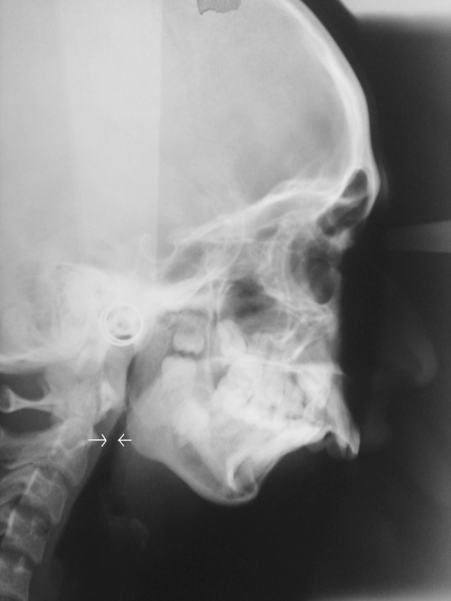
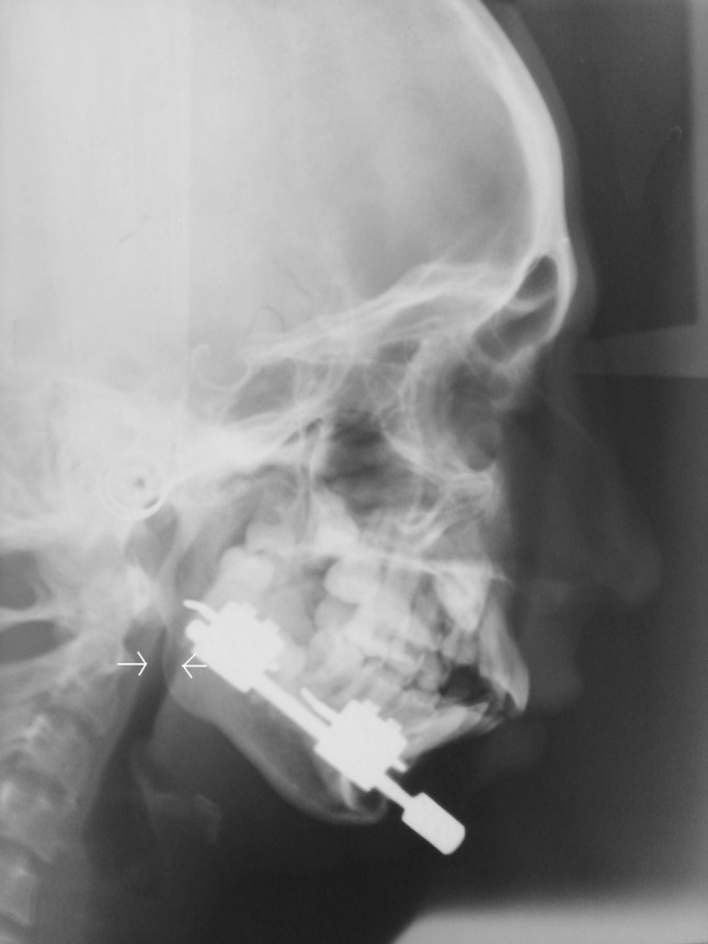
The patients were then evaluated pre distraction and immediate post-consolidation using lateral cephalograms to determine changes in their oro-pharyngeal airway space and dimensions following mandibular distraction. The airway dimensions were first measured using McNamara analysis [5] which utilizes measurements in relation to the upper pharynx and lower pharynx. The upper pharyngeal width is measured from a point on the posterior outline of the soft palate to the closet point on the posterior pharyngeal wall, while the lower pharyngeal width is measured from the intersection of the posterior border of the tongue and the inferior border of the mandible to its corresponding closet point on the posterior pharyngeal wall. The oro-pharyngeal airway space was then traced on the lateral cephalogram using the following radiographic points—Sella (S), Nasion (N), Frankfurt Horizontal (FH), and Pterygomandibular Vertical Plane (PTV) [6]. A line connecting the posterior nasal spine and anterior tubercle of the atlas defined as the superior border of the posterior airway space (PAS), while the line drawn across the median glosso-epiglotic fold parallel to FH defined the inferior border of the PAS. The posterior border of the PAS was defined as the posterior pharyngeal wall (PhW) and the anterior border was defined as the posterior tongue outline and the PTV. The antero-posterior PAS dimension was measured as a line connecting the most posterior point of the tongue (TB) and PhW drawn parallel to FH. The tracings were then super-imposed on a grid and the area involved was manually measured. This provided the approximate change in airway space pre and post distraction.
Surgical Procedure
The surgical procedure in all cases included Linear, single-vector, external distraction for the mandible which was either unilateral or bilateral as indicated. A standard DO protocol of rate 1 mm/day with a rhythm of twice daily and an eight week consolidation was followed. Consolidation was confirmed subjectively with an Orthopantomogram.
Results
The following observations were recorded between the pre-distraction and the immediate post-consolidation values for pulse-oximetry and cephalometry. The mean mandibular advancement in this series was 17.33 mm (Tables 1, 2).
Pulse-oximetry—there was a 3.8 % increase in the oxygen saturation from 91 to 94.8 %. The oxygen desaturation index showed a considerable decrease of 21.62 between the pre and post distraction values.
Cephalometry—a 31.33 % increase in the oro-pharyngeal airspace and an average of 3 mm increase in the upper and lower pharyngeal dimensions were recorded.
Discussion
Micrognathia and retrognathia are common features seen in a number of congenital craniofacial anomalies including Treacher Collins syndrome, Hemifacial Microsomia, Nager syndrome and Pierre robin sequence [3]. Unlike other congenital craniofacial anomalies enumerated above, TMJ ankylosis is an acquired condition which produces a severe degree of retrogenia and micrognathia which may lead to airway compromise and OSAS. OSAS is a condition caused by upper airway obstruction characterized by hypopnoeic-apnoiec episodes during sleep which in extreme conditions may be accompanied by severe secondary cardiac and respiratory problems.
As observed in patients with micrognathia, the additional space occupied by tongue, soft palate and redundant pharyngeal mucosa reduces the cross sectional area of oropharyngeal airway by an average of 25 % [1]. Mandibular distraction provides a consistent change in tongue base position that improves obstructive airway symptoms by increasing measured effective airway space. The use of distraction in the management of hypolastic mandible has shown significant reduction in the respiratory distress index [7]. In our series of cases the mean mandibular advancement with DO was 17.3 mm and the airway changes following distraction obtained by cephalometric analysis showed a mean increase of 31.33 % in airway space, which provided functional rehabilitation by significantly increasing their oro-pharyngeal space and reducing their sleep disturbances as indicated in table.
Various cephalometric studies have demonstrated the effectiveness of mandibular advancement procedures on the improvement in oro-pharyngeal dimensions [7]. Many studies have demonstrated the craniofacial anatomy in OSA using various imaging techniques. Although conventional cephalometry can be done, in supine position, computed tomographic technique carries significant advantages over plain roentgenographic imaging. Considering the cost factor and its application as a mass screening tool cephalometry can be used as a useful diagnostic tool in the analysis of OSA patients.
Cephalometric studies have shown that it is not only the size of these oro pharyngeal soft tissues but also their relative position in the osas patients which adversely affect the integrity of the pharyngeal airway. The mean percentage of change in the airway space was 31.33 % and there was direct change in the oxygen saturation status from 91 to 94.8 %. Though a direct correlation between the improvement of the apnoea/hypopnoea status and the cephalometric changes in the oro-pharyngeal dimensions or the increase in the surface area were not able to be established, there was a directly proportional improvement in the oxygen saturation status with reduction in the ODI4. Despite the limitation of recording a 3D airway on a 2D radiograph the upright lateral cephalogram has been thought to be a useful diagnostic tool in the analysis of snoring and OSA subjects.
Conclusion
The present study highlights the diagnostic as well as prognostic role of cephalometry for clinical perception of geometry of pharyngeal airway in patients having OSAS and also as a mass screening tool along with clinical head and neck examination, polysomnographic and endoscopic studies (Figs. 1, 2).
Fig. 1.

Pre-operative lateral cephalogram demonstrating a severely retrognathic mandible and reduced airway space. Arrows indicate point of maximum constriction
Fig. 2.

Immediate post-consolidation lateral cephalogram with distraction device in situ, demonstrating a significant increase in the airway space. Arrows indicate increase in dimension following distraction
References
- 1.Bond T. Evaluation and diagnosis of sleep-disordered breathing. Oral Maxillofac Surg Clin North Am. 2002;14:293–296. doi: 10.1016/S1042-3699(02)00030-4. [DOI] [PubMed] [Google Scholar]
- 2.Li KK, Guilleminault C, Riley R. Obstructive sleep apnea and maxillo-mandibular advancement: an assessment of airway changes using radiographic and nasopharyngoscopic examinations. J Oral Maxillofac Surg. 2002;60:526–530. doi: 10.1053/joms.2002.31849. [DOI] [PubMed] [Google Scholar]
- 3.Anantanarayanan P, Vivek N, Manikandhan R, Kumar D. Primary mandibular distraction for management of nocturnal desaturations secondary to temporomandibular joint (TMJ) ankylosis. Int J Pediatr Otorhinolaryngol. 2008;72:385–389. doi: 10.1016/j.ijporl.2007.11.015. [DOI] [PubMed] [Google Scholar]
- 4.Saito H, Araki K, Ozawa H, Mizutari K, Inagaki K, Habu N, et al. Pulse-oximetry is useful in determining the indications for adenotonsillectomy in pediatric sleep-disordered breathing. Int J Pediatr Otorhinolaryngol. 2007;71:1–6. doi: 10.1016/j.ijporl.2006.08.009. [DOI] [PubMed] [Google Scholar]
- 5.McNamara JA. A method of cephalometric evaluation. AJO. 1984;86:449–469. doi: 10.1016/s0002-9416(84)90352-x. [DOI] [PubMed] [Google Scholar]
- 6.Tselnik M, Pogrel A. Assessment of the pharyngeal airway space after mandibular setback surgery. J Oral Maxillofac Surg. 2000;58:282–285. doi: 10.1016/S0278-2391(00)90053-3. [DOI] [PubMed] [Google Scholar]
- 7.Arlen DD, Ran T, Pamela HR, René RF. Mandibular distraction osteogenesis in very young patients to correct airway obstruction. Plast Reconstr Surg. 2001;108:302–311. doi: 10.1097/00006534-200108000-00004. [DOI] [PubMed] [Google Scholar]


