Figure 2.
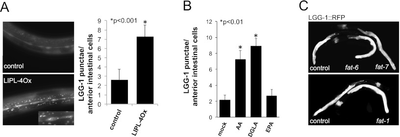
ω-6 PUFAs activate autophagy in C. elegans. (A) LIPL-4Ox activates autophagy. LGG-1∷GFP signal in L3 animals overexpressing LIPL-4 from a constitutively highly expressed intestinal promoter versus control animals is shown (control corresponds to siblings not carrying LIPL-4∷TagRFP but carrying LGG-1∷GFP); exposure time = 100 msec. Quantification of LGG-1∷GFP punctae per anterior intestinal cell from at least three independent experiments is depicted as mean ± SEM using the same exposure time for LIPL-4∷TagRFP and control worms. At longer exposure times (500 msec), more LGG-1∷GFP punctae than at 100 msec can be observed in control animals; for comparison at 500 msec, see Supplemental Figure S5. For bleed-through and autofluorescence controls, see Supplemental Figure S4. (B) AA and DGLA activate autophagy in C. elegans. LGG-1∷GFP punctae per anterior intestinal cell in supplemented (50 μM) or mock-treated (50% ethanol) L3 animals are shown; data from three independent experiments are depicted as mean ± SEM. (C) fat-6, fat-7, or fat-1 inactivation is insufficient to activate autophagy in C. elegans. LGG-1∷RFP worms were egg-laid in fat-1, fat-6, fat-7, or vector control RNAi plates (qPCR analysis showed that the levels of expression of fat-1, fat-6, and fat-7 after RNAi treatment were 0.2 ± 0.1, 0.3 ± 0.05, 0.3 ± 0.1 of vector control treated animals, respectively). The pattern and intensity of the LGG-1∷RFP signal was scored in young adult worms. Representative control and fat RNAi-treated animals are shown. No difference was observed in the intensity or pattern of the expression of LGG-1∷RFP in treated animals when compared with empty vector-treated worms.

