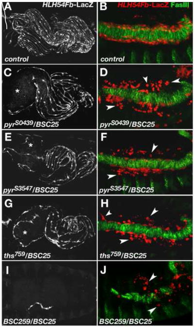
Fig. 3. Abnormal longitudinal visceral muscle development in mutants with reduced FGF ligand activity.
Anti-β-gal staining of HLH54Fb-lacZ embryos to visualize longitudinal visceral muscle fibers at stage 17 (A, C, E, G, I) and of migrating LVM progenitors at the end of germ band retraction (B, D, F, H, J). In the latter panels the TVM is marked by anti-fasciclin III (FasIII) staining in green. (A, B) Wild type. (C) Embryo carrying the EMS-induced mutation pyrS0439 in trans to the pyr- and ths-deleting deficiency Df(2R)BSC25 with a strong depletion of LVM fibers especially around the anterior midgut (asterisk). (D) Late stage 12 embryo of the same genotype as in (C). Numerous LVM founders have lost contact with the TVM and many of them appear as small rounded dots (arrow heads). (E, F) pyrS3547/Df(2R)BSC25 mutant embryo showing essentially the same phenotype as embryos in (C, D). Very similar defects are also observed in ths759/Df(2R)BSC25 mutants (G, H). (I) Removal of pyr and ths using overlapping deficiencies Df(2R)BSC25 and Df(2R)BSC259 (“FGF8 null” genotype) leads to a complete loss of the LVM with rare occurrence of individual LVM fibers. (J) In a FGF8 null mutant, the number of LVM founders at late stage 12 is greatly diminished and almost all cells have lost contact with the TVM.

