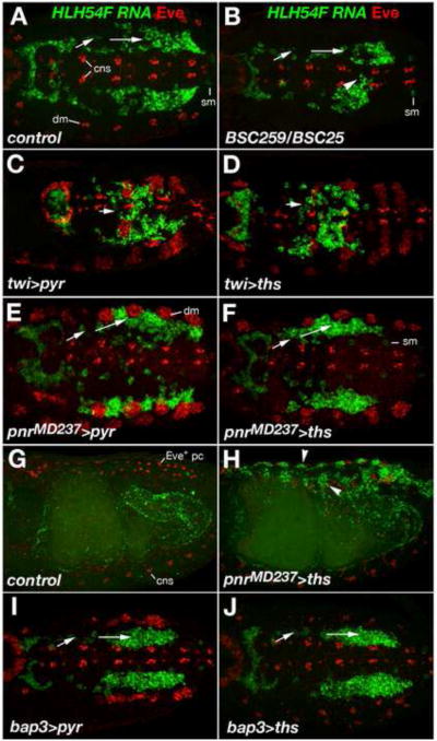
Fig. 8. Effects of Pyr and Ths on the direction of LVM founder migration.
Wild type, mutant, and FGF over-expressing embryos were stained for HLH54F RNA (green) and Even-skipped (Eve) protein (red). In A-F, I, and J, dorsal views of germ band extended embryos are shown with caudal ends to the left and anterior directions toward the right. (A) Stage 11 wild type embryo; HLH54F expression (green) marks migrating LVM founders derived from the caudal visceral mesoderm and Eve (red) marks FGF-dependent precursors of specific pericardial and somatic muscle cells in the dorsal mesoderm (dm). Arrows indicate the lateral-anterior directions of LVM founder migration. HLH54F is also weakly expressed in one ventrolateral somatic muscle progenitor per segment (sm) and Eve also labels cells of the central nervous system (cns; in a lower focal plain than the visceral mesoderm but included in the Z-stack projections as a reference for the location of the ventral midline). (B) FGF8 null mutant stage 11 embryo. At this stage, overall migration is relatively normal but less symmetric, with some cells straying from the normal path and migrating toward the midline and/or the Z direction (arrow head). The number of migrating LVM founders is slightly reduced. There is no eve activation in the dorsal mesoderm. (C, D) Ectopic expression of either pyr (C) or ths (D) in the entire mesoderm via twi-GAL4 (SG24) prevents LVM founders from forming bilateral groups. Migration of individual cells appears to be undirected with only little net movement toward the anterior trunk. Mesodermal Eve clusters are enlarged. (E, F) Ectopic expression of either pyr (E) or ths (F) in the dorsal ectoderm via pnrMD237-GAL4 causes LVM founders to migrate further laterally as compared to cells of the same stage in normal embryos. LVM founders between the (enlarged) Eve clusters of the dorsal mesoderm extend processes toward the ectoderm. (G) Lateral view of stage 15 wild type embryo in which LVM fibers have aligned along the entire midgut. Eve expression is seen dorsally in Eve+ pericardial cells, weakly in somatic muscle DA1, and ventrally in the CNS. (H) Stage 15 embryo with pnrMD237-GAL4-driven ectopic expression of ths in the dorsal ectoderm. LVM fibers are seen along the entire midgut as in (G), but many LVM cells have been redirected toward dorsal areas underneath the ectoderm (arrow heads). (I, J) LVM founders migrate normally in stage 11 embryos when either pyr (I) or ths (J) are over-expressed in the TVM via bap3-GAL4 (compare to A).

