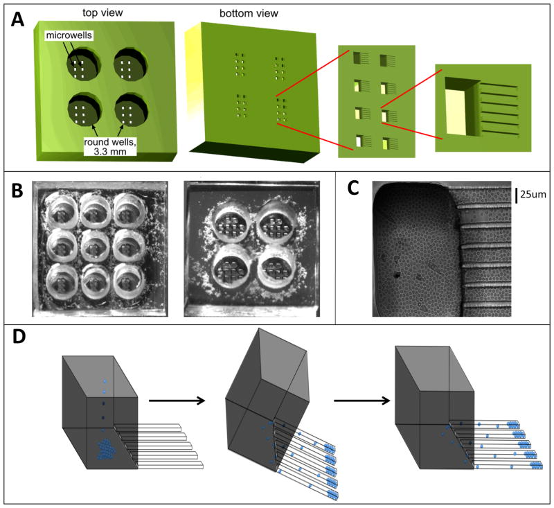
Figure 1. The microfabricated device with finger-like microchannels.
(A) Schematic of a PDMS chip with a 2×2 array of 3.3 mm in diameter, 3-mm-deep round wells. At the bottom of each round well there is a 2×4 array of 100×200-μm rectangular openings in a 100-μm-thick layer of PDMS, forming microwells. At the bottom of each microwell there are finger-like horizontal extensions, 25×25 μm in cross-section and ~100–200 μm in length, forming cell-imaging channels. (B). Photographs of PDMS chips with 3×3 and 2×2 arrays of round wells. The microwells are visible at the bottom of the round wells. (C) Brightfield image of resting primary CD4+ T lymphocytes loaded into a microwell and the adjacent finger-like channels. (D) Schematic of loading of cells into finger-like channels. First, cells are loaded into a round well and allowed to settle onto the bottoms of the microwells by gravity. Then, the device is tilted by ~45° with the finger-like channels pointing downward, making cells slide into the finger-like channels. Third, the normal orientation of the device is restored and it is carefully placed on the microscope stage.

