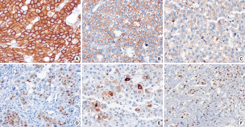
Fig. 4.
Immunohistochemistry of the three cases. The tumor cells tend to be positive for cytokeratin 18 (A, case 1), E-cadherin (B, case 1), epidermal growth factor receptor (C, case 1), and p53 (D, case 2). (E) Serum human chorionic gonadotropin is focal positive (case 3). (F) MIB-1 labeling index of case 1 is 36.1%.

