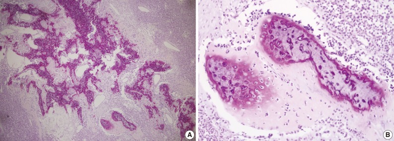
Fig. 3.

(A) Areas of osseous matrix with calcification and partly well-formed bony trabeculae are observed. (B) The periphery of osteoid and bone formation shows frankly malignant tumor cells, but the central portion of the bone reveals lacuna containing, benign-looking nuclei.
