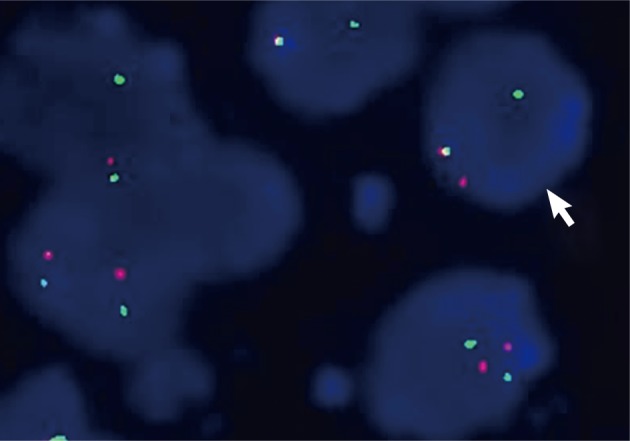
Fig. 5.

Ewing sarcoma breakpoint region 1 (EWSR1) fluorescence in situ hybridization (FISH) on interphase cells showing split-apart signals. Interphase nuclei with fused orange and green hybridization signals are interpreted as indicative of an intact (not rearranged) copy of the EWSR1 gene. A split signal pattern (one green and one orange) seen on interphase nuclei is interpreted as indicative of a EWSR1 gene rearrangement. This case has evidence of EWSR1 rearrangement by FISH.
