
Fig. 2.
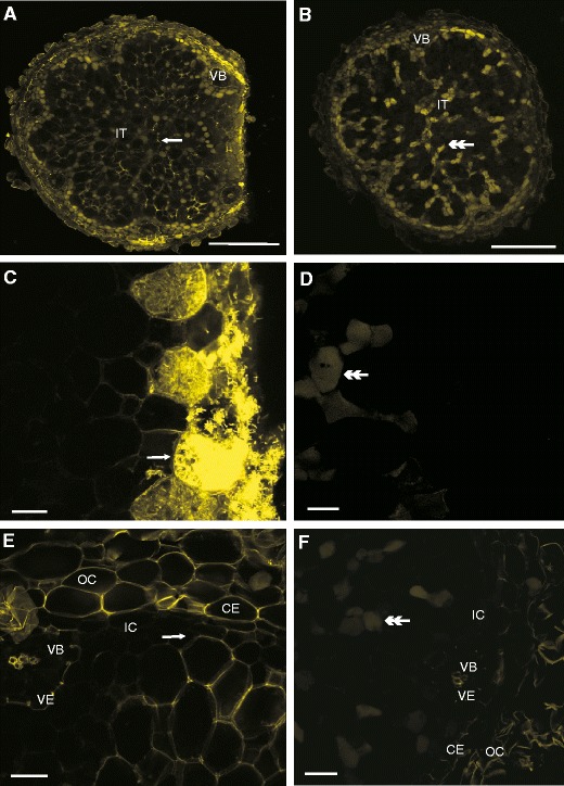
The CLSM photographs of non-fixed sections. Spreading of LYCH through damaged root nodule (a,c,e) with a comparison to a control nodule inner tissue (b,d,e). Notice the dye within the cell walls and the lack of fluorescence in the inner cortex cell walls (e). The cells adjacent to the place of cutting are strongly fluorescent (c). The control root nodule exhibits only pale yellow autofluorescence of uninfected cells (b,d,f). Abbreviations:, CE – common endodermis, IC – inner cortex, IT – inner tissue, OC – outer cortex, VE – vascular endodermis, VB – vascular bundle, arrows – parts of cells stained with LYCH, double arrow-head – autofluorescence of uninfected cells. Bars represent: A and B = 300 μm, C, D, E and F = 20 μm