
Fig. 3.
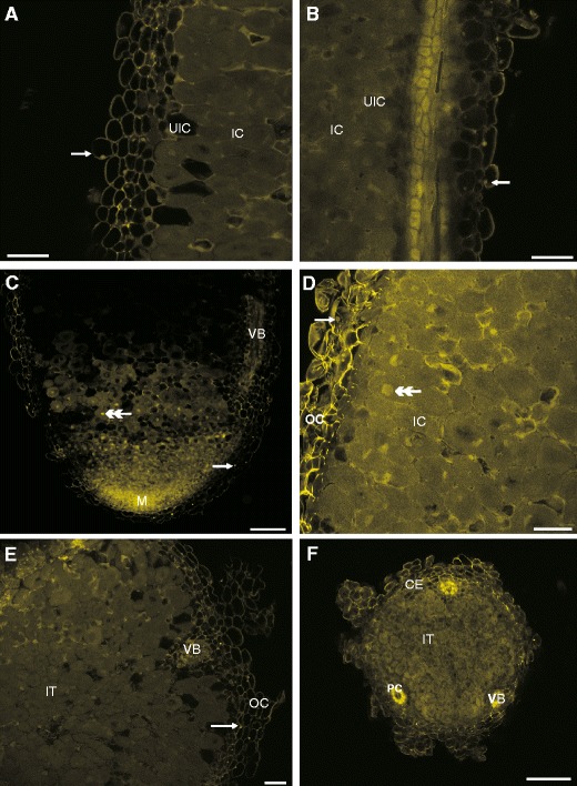
The CLSM photographs of non-fixed root nodules stained with LYCH (a, c, e) and the influence of glutaraldehyde fixation on the fluorescence properties of root nodule staining with LYCH and subsequently fixing in glutaraldehyde (b, d, f). Notice fluorescence of the nuclei (arrows in a vs. b, c vs. d, e), the vacuoles (c vs. d), outer cortex (a vs. b, c vs. d, e vs. f) and pericycle of vascular bundle (f). Abbreviations:, CE – common endodermis, IC – inner cortex, IT – inner tissue, M – meristem, OC – outer cortex, PC – pericycle cells, UIC – uninfected cells, VB – vascular bundle, arrows – parts of cells stained with LYCH, double arrow-head – autofluorescence of uninfected cells. Bars represent: A and B = 60 μm, C = 100 μm, D and E = 30 μm, F = 200 μm