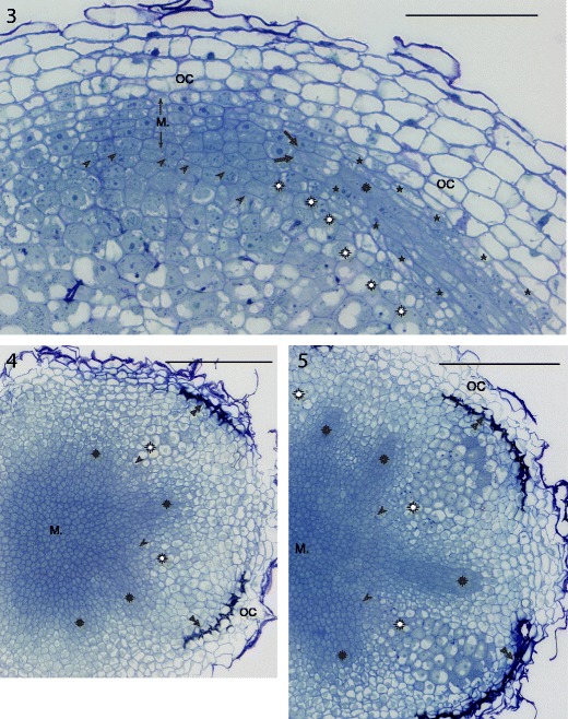
Figs. 3, 4, 5.

Provascular meristems (rosettes) are an integral part of the nodule meristem (M). Fig. 3: Astragalus sinicus root nodule apex. Asterisks — vascular endodermis layer, large arrows — founder cells of provascular meristem. Fig. 4, 5: two transversal sections of the same nodule of Trifolium repens, the second one is cut close to the proximal face of the nodule meristem. Double arrowheads — layer of differentiated cortical endodermis. Common labels: OC — outer cortex, arrowheads — infection threads in the cells adjoining nodule meristem, white rosettes — differentiating cells of bacteroid-containing tissue. Semithin sections stained with azure a and methylene blue, bright field photograph. Bars represent 100, 300, and 300 μm, respectively
