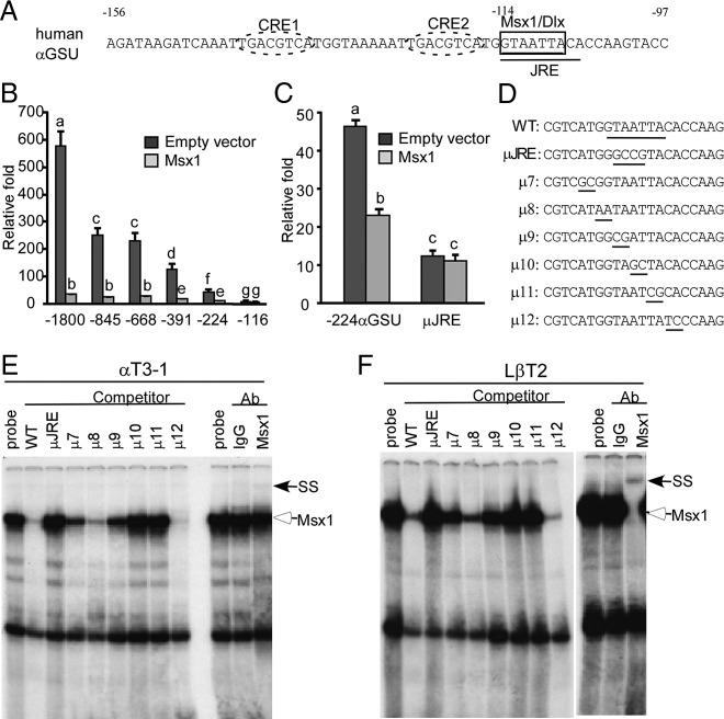
Figure 3.
Msx1 represses the αGSU promoter at the −114 JRE within −224 bp of αGSU mRNA start site. (A) Gonadotrope regulatory elements in the proximal human αGSU promoter. Thick boxes indicate the putative Msx1/Dlx-binding elements, the −114 site. The JRE element is underlined. CRE1 and CRE2 are marked with dashed ovals. (B) The full-length human αGSU promoter and its truncated reporters map Msx1 repression inside of −224 bp of the start site on the αGSU promoter. The wild-type −1800 αGSU-luc, and its truncations: −845 αGSU-luc, −668 αGSU-luc, −391 αGSU-luc, −224 αGSU-luc, and −116 αGSU-luc, were transiently transfected into αT3-1 cells with the Msx1 expression vector or an equal mass of empty expression vector. (C) Identification of Msx1-binding sites on the human αGSU promoter. Wild-type −224 αGSU reporter (WT) and mutants (μJRE) were transiently transfected into αT3-1 cells, pGL3 empty luciferase reporters were used as control. Cells were cotransfected with Msx1 or an equal mass of empty expression vector. Groups labeled with different letters are significantly different from each other (P < .05). (D) The oligonucleotides used for the αGSU JRE probe and its mutants (μ7–μ12) are listed, and the Msx1-binding element and 2-bp transversions are underlined. Msx1 binding on the αGSU promoter maps to the −114 site in αT3-1 (E) and LβT2 (F) gonadotropes. The hollow arrow marks the Msx1 complex, and the arrow marks the supershift of the Msx1 complex after binding with Msx1 antibody. SS represents supershift complex. Abbreviation: Ab, antibody.

