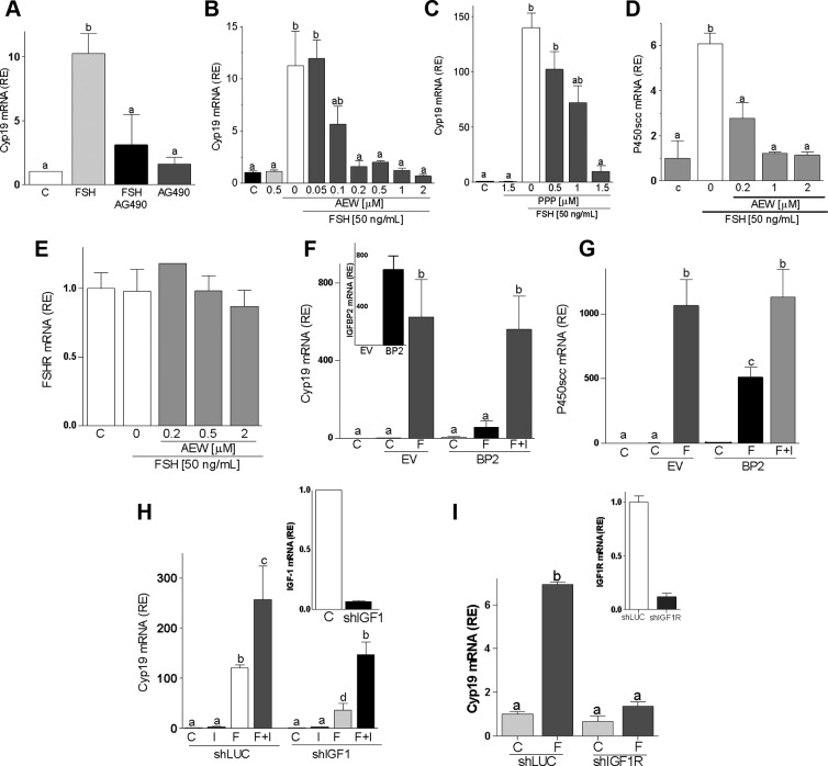
Figure 3.
Inhibition of IGF-IR activation blocks FSH-induced differentiation. Rat GCs were cultured with FSH plus AG490 (25μM, panel A) or different concentrations of AEW (panels B, D, and E) or PPP (panel C). mRNA levels for Cyp19 (panels A–C), P450scc (panel D), or FSHR (panel E) were determined after 48 hours of treatment. Panels F and G, GCs transfected with a plasmid expressing IGFBP2 (BP2) or the empty vector (EV) were treated with FSH (50 ng/ml) or vehicle for 48 hours before Cyp19 and P450scc mRNA level quantification. The inset in panel F shows IGFBP2 expression levels in cells transfected with EV or BP2. Panels H and I, GCs were infected with a lentivirus carrying a control shRNA (shLUC or C), anti–IGF-I shRNA (shIGF-I), or anti–IGF-IR shRNA (shIGF-IR). At 24 hours after infection, cells were treated with FSH (50 ng/ml) and/or IGF-I (50 ng/ml), and Cyp19 mRNA levels were quantified 48 hours later. The inset shows IGF-I (panel H) or IGF-IR (panel I) mRNA levels in cells infected with shLUC, shIGF-I, or shIGF-IR. Each experiment was performed three times, and mean and SEM are shown. Columns with different letters differ significantly (a-b and b-c P < .05; a-c P < .01).

