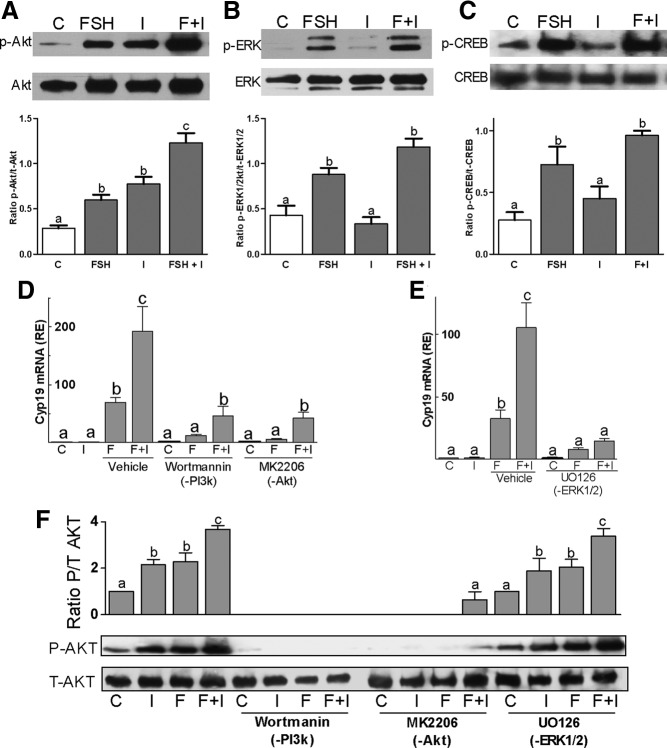
Figure 6.
FSH and IGF-I signaling cross talk. Rat primary GCs were treated with FSH (50 ng/ml) and/or IGF-I (50 ng/ml) for 1 hour. A–C, Phosphorylated and total forms of AKT (panel A), ERK1/2 (panel B), and CREB (panel C) levels were determined by Western blot. The bar graphs under each blot show the mean ± SEM of the ratio of phosphorylated to total protein of three or more experiments. Panels D and E, Rat GCs were pretreated with wortmannin (100nM), an inhibitor of PI3K; MK2206 (1μM), an inhibitor of AKT; or UO126 (5μM), which inhibits ERK1/2, for 1 hour followed by treatment with FSH and/or IGF-I for 48 hours. Cyp19 levels were quantified by real-time PCR. Panel F, GCs were pretreated for 1 hour with wortmannin, MK2206, or UO126 followed by 1 hour treatment with vehicle or FSH and/or IGF-I before protein isolation. Phosphorylated and total AKT protein levels were evaluated by Western blot. Each experiment was repeated 6 times. Columns with different letters differ significantly (a-b and b-c P < .05; a-c P < .01). Abbreviations: C, control; F, FSH; I, IGF-I; p, phospho.

