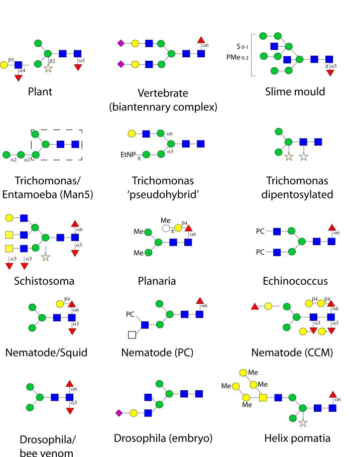
Figure 2. Examples of N-glycan structures from a selection of non-vertebrate eukaryotes.
In comparison to plants and vertebrates, examples of N-glycans from Dictyostelium discoideum (slime mould), Trichomonas vaginalis (protozoal parasite; the ‘biosynthetic’ Man5 structure being found also in Entamoeba histolytica, with the trimannosylchitobiosyl region being boxed with a dashed line), Schistosoma mansoni (trematode parasite), Dugesia japonica (planaria), Echinococcus granulosus (cestode parasite), Caenorhabditis elegans (nematode; the ‘GalFuc’ epitope being also found in some molluscs), Drosophila melanogaster (fruitfly; difucosylation also being found on bee venom glycoproteins) and Helix pomatia (mollusc) are shown. Incomplete lines indicate further structural possibilities. CCM core chitobiose modification, EtNP indicates ethanolamine phosphate, Me methyl, PC phosphorylcholine, PMe methylphosphate, S sulphate. Monosaccharides are depicted according to the nomenclature of the Consortium for Functional Glycomics (see Figure 1).

