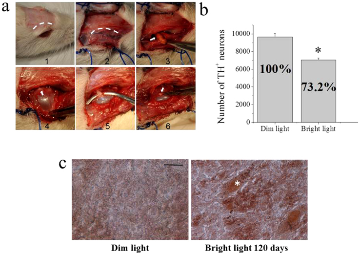
Figure 5. TH immunohistochemistry and stereological counting of TH-positive neurons in ventral mesencephalon of the bilateral optic nerve transected rats.
(a) Surgical transection of the optic nerve. In panel 1 and 2 the curvilinear dashed lines indicate the incision of the scalp and the incision along the orbital rim to expose the retro-orbital tissue, respectively. Excess of retro-orbital fat (indicated by the arrow in panel 3) was removed and the orbit pulled forward to expose the optic nerve (indicated by the arrow in panel 4). The optic nerve was hooked with a surgical instrument (panel 5) and then cut. The arrow in panel 6 indicates the stump of the transected optic nerve. (b) Stereological counting of TH-positive neurons in the substantia nigra of rats raised in dim light – dark cycle, and rats raised under bright light for 4 months. *Significantly different from animals raised in dim light – dark cycle (t8,8 = 5.87463, p = 0.00004, using two-samples t-Test). (c) Fontana-Masson staining of neuromelanin granules in the substantia nigra. Representative sections from rats raised in dim light – dark cycle (left panel) and rats raised under bright light (right panel). The asterisk on the right panel shows a cell with numerous tiny neuromelanin granules.

