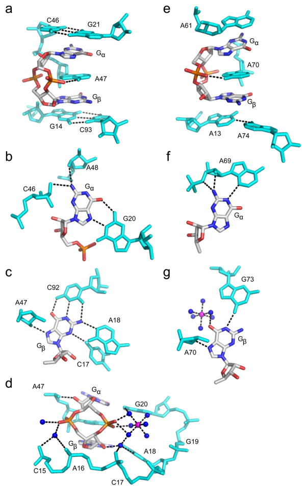
Figure 4.
RNA recognition of c-di-GMP. c-di-GMP is colored by atom as in Figure 3. RNA residues are colored cyan, water molecules are shown as blue spheres and magnesium ions as magenta spheres. Hydrogen bonds are indicated by black dashed lines. (a) c-di-GMP bound to the Vc2 class I riboswitch from Vibrio cholerae (PDB ID 3MXH). (b) Recognition of Gα,(c) Gβ and (d) the c-di-GMP ribosyl-phosphate backbone by the class I riboswitch. (e) c-di-GMP bound to the Cac-1-2 class II ribowitch from Clostridium acetobutylicum (PDB ID 3Q3Z). (f) Recognition of Gα and (g) Gβ by the class II riboswitch.

