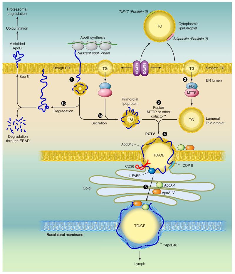
FIGURE 3.
Model of intestinal triglyceride-rich lipoprotein assembly. The nascent apoB polypeptide is cotranslationally translocated across the rough endoplasmic reticulum (ER) membrane (1). The membranes of the ER are shown in yellow. When lipid (triglyceride, TG) is available, physical interactions between the NH2-terminal domain of apoB and the microsomal triglyceride transfer protein (MTTP) promote optimal folding and direct biogenesis of a primordial lipoprotein particle (1a). MTTP exists as a heterodimeric complex with the chaperone protein protein disulfide isomerase (PDI). MTTP also promotes mobilization of triglyceride-rich lipid droplets from the bilayer of the adjacent smooth ER into the lumen of the ER to become luminal lipid droplets (2). When lipid availability is limited or MTTP function is impaired, nascent apoB becomes misfolded (1b) and is degraded, either within the ER lumen via ER-associated degradation pathways (ERAD), or the misfolded protein undergoes ubiquitination and is degraded by the proteasomal pathway. During the second step of lipoprotein assembly, the primordial lipoprotein particle fuses with endoluminal lipid droplets, resulting in prechylomicron transport vesicle (PCTV) formation (3). Other proteins, including CD36 and L-Fabp, also participate in PCTV formation. After fusion with key vesicular transport proteins, including COP II proteins, the prechylomicron particles are incorporated into a vesicular complex that buds from the ER and fuses with Golgi membranes. Vectorial transport of these prechylomicron particles (4) results in their continued maturation and eventually secretion (5) of the mature, nascent chylomicron particles into the pericellular spaces adjacent to lymphatic fenestrae. The basolateral membrane (shown schematically with a mature chylomicron traversing into the lymphatic fenestrae) is illustrated in blue.

