. 2013 Mar 5;2:e00327. doi: 10.7554/eLife.00327

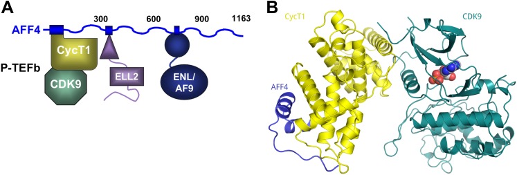
Figure 1. AFF4 binds CycT1 distal to CDK9.

(A) Schematic model of the SEC. AFF4 is an intrinsically disordered scaffold that binds partners via 20–50 residue segments. (B) Ribbon diagram showing the strand–helix–helix arrangement of AFF4 (blue) bound to CycT1 (yellow) remote from CDK9 (teal). AFF4 adopts an extended conformation with no intramolecular tertiary contacts. AMPPNP (spheres) is bound to CDK9.

DOI: http://dx.doi.org/10.7554/eLife.00327.003

Figure 1.

Figure 1—figure supplement 1. Electron density for AFF42–73.
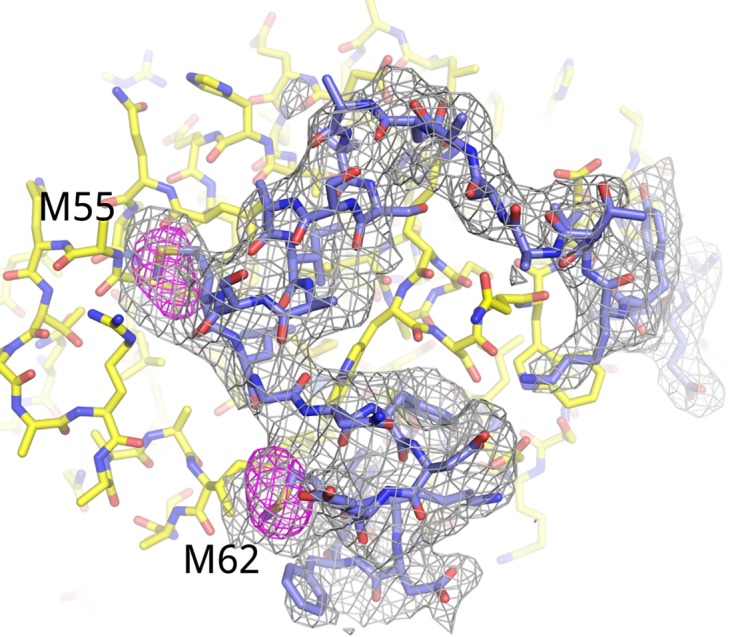
Figure 1—figure supplement 1.

Fo-Fc omit map (3.5 σ, gray) for residues 34–66 of AFF4. Anomalous difference map (5 σ, red) shows the positions of the methionine residues in AFF4. The anomalous difference Fourier was calculated using X-ray data recorded from a crystal grown with Seleno-methionine labeled AFF4.

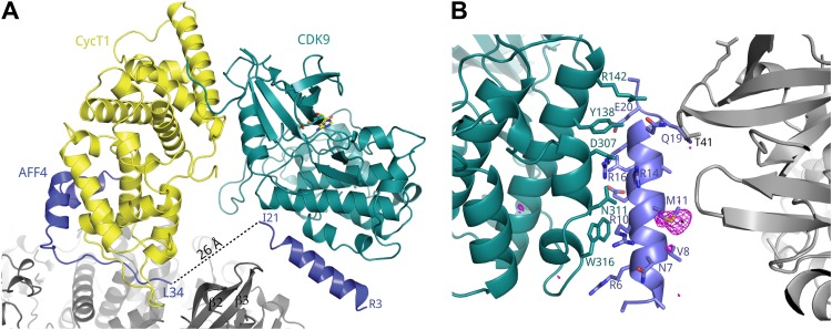
Figure 1—figure supplement 2. A crystal contact formed by AFF42–21.

Figure 1—figure supplement 2.

(A) An isolated helix from the aff4 N-terminus, packs loosely against αE and αI of one CDK9 subunit (chain C) and makes contacts with the β2-β3 loop of a symmetry-related CDK9 molecule. (B) Interactions between CDK9 residues (teal sticks) and the isolated aff4 helix H0 (blue). Anomalous difference map (3.5 σ, red) shows the position of SeMet11. The occurrence of this helix in only one of three independent complexes in the crystals and the lack of electrostatic complementarity with CDK9 suggest that the helix may depend on the crystal environment and the kinase subunit is not the functional partner of this sequence.