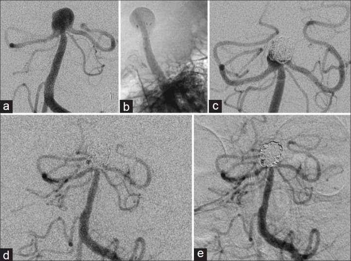
Figure 3.

Patient 1 cerebral angiogram (left vertebral injection). (a) AP view showing a basilar apex aneurysm pretreatment. (b) Lateral view of the Enterprise stent deployed in the aneurysm dome. (c) Follow-up AP view showing small basilar apex aneurysm recurrence before redo treatment. (d) Follow-up AP view after redo coil embolization of recurrent aneurysm. (e) 27-month follow-up AP view after redo coil embolization showing no further recurrence
