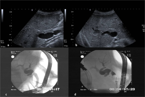
Figure 1. The Images Show a Stenosis at The Level of The Anastomosis With Intrahepatic Dilatation of The Bile Ducts.

Figure 1a demonstrates a liver recipient with a biliodigestive anastomosis in a longitudinal position of the ultrasound probe in the media clavicular line. Dilatated gut loop with fluid in liver hilum. Figure 1b demonstrates the right lobe of the same patient in a sub costal view. Dilatated right common bile duct with a peripheral abscess (arrow) due to the stenosis of the bile duct at the level of the bilio digestive anastomosis. Figure 1c and 1d demonstrate the corresponding ERCP images without and with blocked balloon catheter
