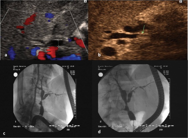
Figure 2. The Images Show Ischemic Type Biliary Lesions (ITBL) Signs at the Level of The Anastomosis as Well as The Intrahepatic Bile Ducts.

Figure 2a demonstrates a centrally dilated right common bile duct of a liver recipient in color mode. Figure 2b demonstrates the same patient with partially thickening of the wall (arrow) of the common right bile duct representing an ischemic biliary type lesion. Figure 2c and 2d demonstrate the corresponding ERCP images without and with blocked balloon catheter
